Microscopy images are only as powerful as the analysis behind them. On Friday, March 27, 2026, the Core Facility Microscopy at the Biozentrum of Martin Luther University Halle-Wittenberg hosted a full-day beginner workshop on ImageJ/Fiji — one of the most widely used tools for scientific image analysis in the life sciences.
The free, hands-on course guided participants through the essentials of quantitative bioimage analysis: from understanding the nature of digital images to segmenting and measuring objects, running particle analyses, and taking first steps in automating repetitive tasks. All with their own laptops, their own data — and the support of four experienced instructors.
Topics covered included:
- The basics of digital images and common file formats
- Transforming raw microscopy data into publication-ready figures
- Placing scale bars and extracting histograms
- Segmenting and measuring objects
- Particle analysis
- Working with ImageJ plugins
- An introduction to task automation

The workshop was organised by Dr. Martin Schattat, Dr. Birgit Möller, Dr. Markus Glaß, and Dr. Stephanie Krüger — bringing together expertise from plant physiology, computer science, molecular medicine, and the Core Facility Microscopy.
We would also like to extend a warm thank you to RTG 2498 and AgriExplore for kindly supporting the workshop with snacks and drinks — their contribution made the day even more enjoyable for all participants.
Stay tuned for future workshops and events from the Core Facility Microscopy.
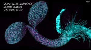
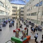
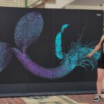
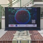
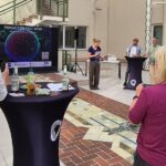
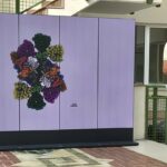
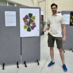
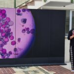
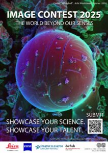
 Scientists, researchers, and students from Martin Luther University and regional institutions are invited to submit their best scientific images — from microscopy to data visualizations. Selected entries will be displayed in a four-week public exhibition, culminating in a live pitch night at the planetarium.
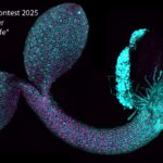
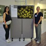
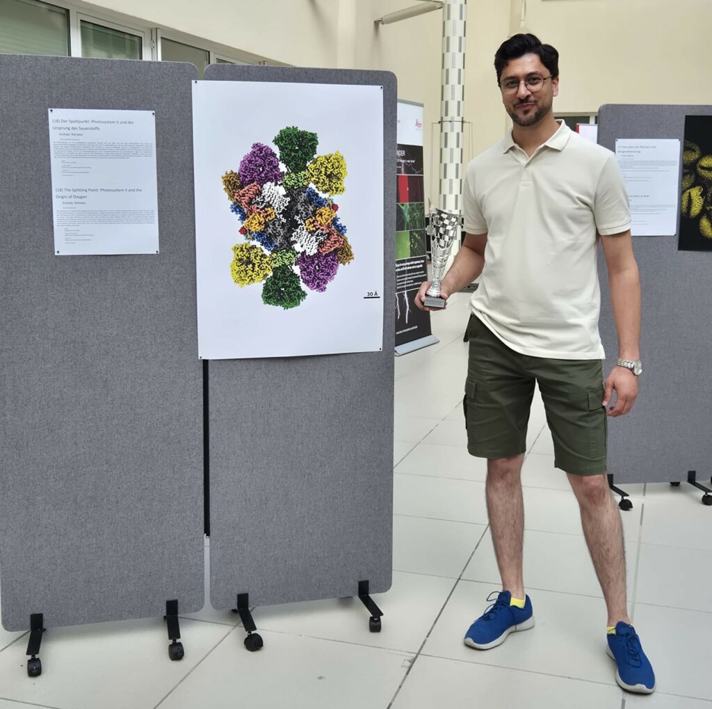
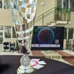
 We look back on a successful year. In addition to a winning image competition and the switch to a digital laboratory management and archiving system, we can look back on 48 processed sample systems from 18 cooperation partners and 11 publications.