Our paper on the 2.7 Å structure of apoferritin with the Glacios and the Falcon 3EC is available as a preprint!

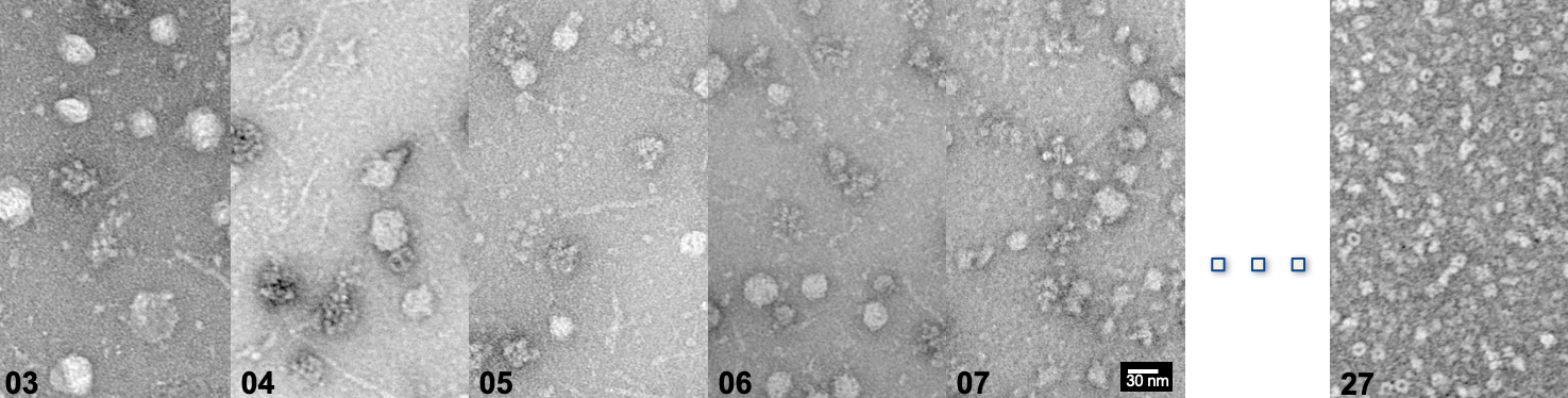
Our paper is available as a pre-print in Biorxiv (https://www.biorxiv.org/content/10.1101/738724v1). In this work, we have determined the atomic structure of mouse apoferritin using the Glacios microscope. Its difference comparedd to other 200 keV microscope is its compact design, allowing the Continue reading Our paper on the 2.7 Å structure of apoferritin with the Glacios and the Falcon 3EC is available as a preprint!

Congratulations to Dmitry – Awarded accommodation fellowship for cryo-EM course

Besides relion, which we currently use for image processing, scipion is another software to analyze images and reconstruct electron density maps and/or calculate cryo-EM models. We would like to explore cryo-EM image processing with scipion, as it is a software Continue reading Congratulations to Dmitry – Awarded accommodation fellowship for cryo-EM course

The cover for Biol Chem – Structural Biology of Native Cell Extracts

We are very proud for our cover for Biol Chem, July issue – it is currently online, and advertised on the main page of the journal. A high-resolution pdf file with explanation of our cover can be found on the Continue reading The cover for Biol Chem – Structural Biology of Native Cell Extracts

Vitrified apoferritin in Halle – first overnight acquisition is complete!

We have successfully vitrified apoferritin in own Vitrobot Mark 4. Loading the microscope and starting EPU yesterday evening ended up with an acquisition of ~300 high-resolution images from a single gridsquare! First of all, we are able to demonstrate the Continue reading Vitrified apoferritin in Halle – first overnight acquisition is complete!